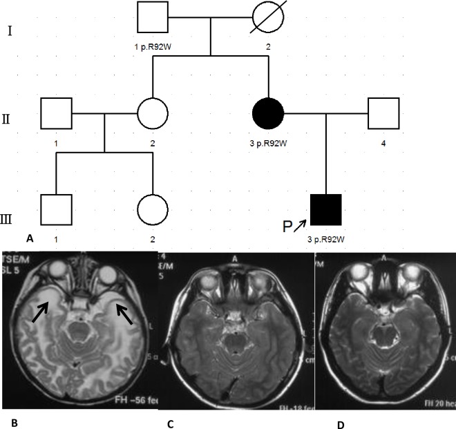
Fig 2. Genogram and cranial MRI of Pt17 and his mother.
(A) Genogram of Pt17. (B) MRI of Pt17 at 7mo. Abnormal signals in the white matter with subcortical cysts in the temporal lobes (Arrows). (C) MRI of Pt17 at 7.67yo. Normal signals in the white matter without subcortical cysts in the temporal lobes. (D) MRI of his mother at 33yo. Normal signals in the white matter without subcortical cysts in the temporal lobes.

