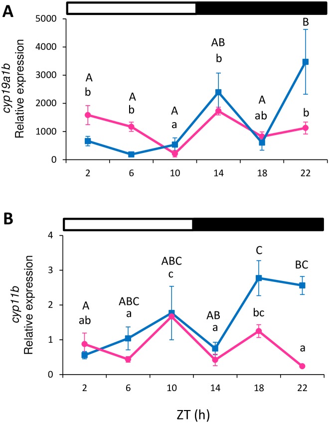
Fig 5. Relative gene expression of cyp19a1b (A) and cyp11b (B) in the brain.
Brain samples were analyzed separately for females (pink circles) and males (blue squares). Data are expressed as mean±SEM. Different letters indicate the statistically significant differences between time points in the female (lower case letters) or male (capital letters) brains (one-way ANOVA, p<0.05) within each gene. The white and black bars above the graphs represent the light and dark phase, respectively.

