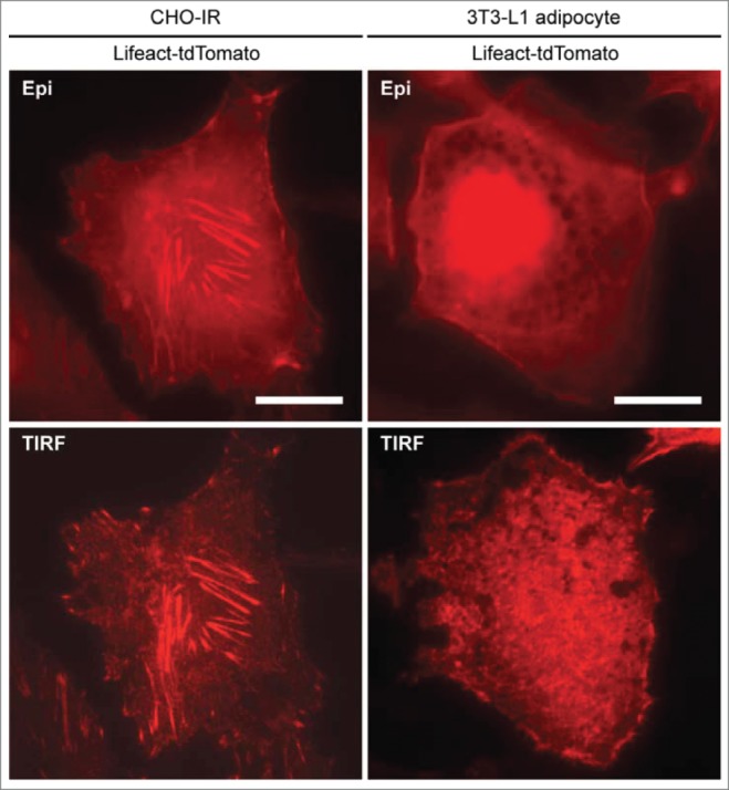
Figure 1.

Comparison of actin structures between CHO-IR and 3T3-L1 adipocytes. CHO-IR (CHO cells stably expressing insulin receptor) and 3T3-L1 cells were imaged by using epifluorescence (Epi) or total internal reflection fluorescence (TIRF) microscopy. Cells expressing Lifeact-tdTomato were seeded in 35 mm glass-bottom dishes (MatTEK), incubated in imaging buffer and kept in a Tokai Hit temperature controlled incubation box at 37°C supplemented with 5% CO2. TIRFM setup (TIRF laser 561 nm) was based on Nikon Eclipse-Ti inverted microscope with EMCCD camera (1002 × 1002 pixels, 8 × 8 μm, 14-bit, Andor iXonEM + 885; Andor Technologies). Images were captured using a 100x NA/1.49 APO TIRF oil-immersion objective with immersion oil (nd = 1.515, Nikon) bridging the optical contact between the objective and the glass bottom dishes. The penetration depth of the evanescent field is estimated to be ∼200 nm. Images were acquired with no binning, at 27 MHz readout rate with average exposure times vary between 50∼100 ms. As revealed in TIRF microscopy, abundant stress fibers are present in the ventral section of the CHO-IR cell whereas cortical actin and short ventral actin filaments are enriched in the adipocyte. Scale bars, 20 μm.
