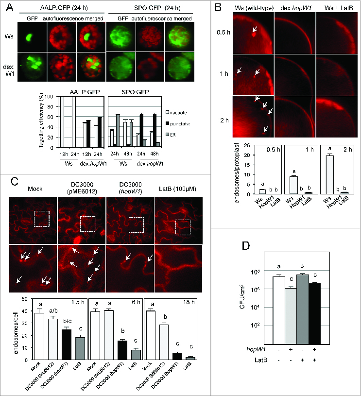
Figure 1 (See previous page).

Effects of HopW1 on actin dependent processes and bacterial growth in Arabidospsis Ws. (A) HopW1 inhibits vacuolar and ER trafficking. Wild-type and dex:hopW1 protoplasts were transfected with AALP:GFP or SPO:GFP, incubated in 0.2 μM of dexamethasone for indicated times and imaged by confocal microscopy. AALP:GFP was targeted to the central vacuole and SPO:GFP localized to the ER and vacuole in wild-type protoplasts (upper row), as previously documented for Col.12 In the presence of HopW1, many protoplasts transfected with the AALP:GFP and SPO:GFP showed notable punctate patterns. Localization patterns were quantified from at least 100 images, see 12 for comparison with Col protoplasts and the effect of LatB. Bars indicate SEM. χ2 tests indicated that the distributions were significantly different between the wild-type and dex:hopW1 at each time point (P < 0.0001, n ≥ 100 per genotype/fusion construct). (B) HopW1 inhibits endocytosis in protoplastas. Representative microscopic images show effects of HopW1 and LatB on endocytic vesicle formation. Wild-type and dex:hopW1 protoplasts were treated with 0.2 μM dexamethasone, stained with FM4–64 and visualized by confocal microscopy. Ten μM LatB was used to disrupt the actin cytoskeleton. After over-night incubation with dexamethasone and/or LatB, protoplasts were labeled with FM4–64 and viewed after 0.5, 1 and 2 h. Arrows point to some of the FM4–64-stained endosomes. Endosomes were quantified in at least 20 protoplasts per treatment, per time-point in 3 independent experiments. Bars indicate SEM, and letters indicate significantly different number of endosomes (P < 0.0001, ANOVA/Neuman-Keuls test). (C) HopW1 inhibits endocytosis during infection. Cotyledons of Arabidopsis Ws seedlings grown on MS plates were infected with PtoDC3000 carrying either empty vector (pME6012) or vector with the HopW1 gene at OD600 = 0.01. 100 μM LatB was used as an actin cytoskeleton-disrupting control. After infections and treatments for the indicated times, cotyledons were labeled for 1 h with FM4–64 and viewed. Examples of microscopic images of tissue 6h after infection are shown. Lower row is magnification of fragments from pictures in upper row. Arrows indicate some of the FM4–64-labeled endosomes. Enodosomes per cell were manually counted in at least 8 images per treatment, per timepoint, from 2 biological repeats. Bars indicate SEM and letters indicate significantly different numbers of endosomes for given treatments (P < 0.05, ANOVA/Neuman-Keuls test). (D) Disruption of actin cytoskeleton does not induce resistance in Arabidopsis Ws. Growth of PtoDC3000/vector or PtoDC3000/HopW1 (OD600 = 0.0001) 3 d post-inoculation of Ws accessions was monitored in the presence or absence of 10 μM LatB. Different letters indicate significantly different growth (ANOVA/Neuman-Keuls test, P < 0.05). LatB did not trigger resistance in Ws, whereas HopW1 did, suggesting that the mechanism of resistance does not involve disruption of the actin cytoskeleton. Average of all results with SEM from 3 independent experiments (n=9) is shown. All experiments were repeated 3 times with similar results. Detailed methods are described in ref.12
