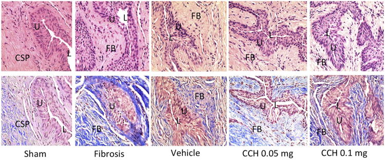
Figure 1.

Histology of rat urethral tissue sections (top-H&E stain, bottom-MT stain-Bottom, ×400) demonstrating normal urethral lumen surrounded by normal distribution of collagen bundles and smooth muscle cells without fibrosis in the sham group, moderate fibrosis with densely packed collagenous stroma involving submucosal tissue in the urethral fibrosis group, mild submucosal fibrosis in the vehicle group, mild submucosal fibrosis in the low-dose CCH group, and minimal fibrosis in the high-dose CCH group. CSP, Corpus spongiosum; FB, fibrosis; L, Lumen; U, urothelium.
