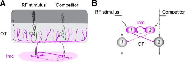
Figure 4. Anatomically grounded circuit model.
(A) Schematic of anatomical connectivity between the OT and Imc (Goddard et al., 2014; Wang et al., 2004). Shaded circles: OT neurons; purple ovals: Imc neurons. Two spatial channels (#1 and #2) are shown; one of them is represented with dashed lines for visual clarity. (B) Schematic of model circuit that respects the anatomy. Shaded circles: OT units; purple ovals: Imc units. Arrows: excitatory connections; lines with spherical heads: inhibitory connections.

