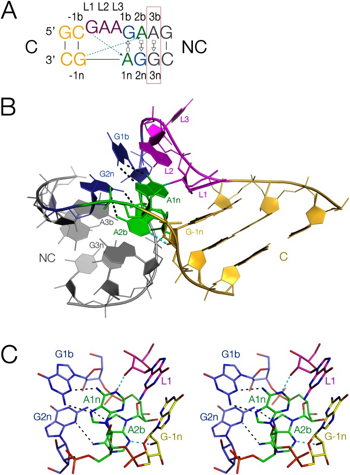
Figure 1.
Standard k-turn sequence and structure. (A) The sequence of Haloarcular marismortui Kt-7, with the nucleotide positions labeled using the established nomenclature (1). The 3b,3n basepair studied here is boxed red. The two key cross-strand hydrogen bonds are shown as broken arrows colored cyan. (B) The structure of HmKt-7. The coloring matches that of the sequence in part A. The bulged strand is at the back in this view, with the non-canonical helix (NC) on the left, and the canonical helix (C) on the right. (C) The A-minor hydrogen bonds (broken lines) in the core of the k-turn structure shown in parallel-eye stereoscopic view. The two cross-strand hydrogen bonds are highlighted cyan.

