Figure 3.
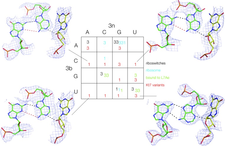
The conformation of HmKt-7 3b,3n-variants within the SAM-I riboswitch context. The structures of 12/16 variants have been determined from which we have deduced the N3 (3) or N1 (1) conformation (see also Supplementary Figure S3). These are shown in red within the 4 × 4 array, added to those of the natural k-turns colored as in Figure 2. The structures of the 2b•2n•-1n triple base interactions are shown for four of the modified Kt-7 k-turns. Hydrogen bonds are indicated by black broken lines. Red broken lines indicate distances too long to be hydrogen bonded. The complete set of 2b•2n•-1n triple base structures is presented in Supplementary Figure S4.

