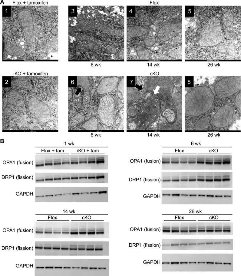
Figure 4.
Morphology and biochemical characteristics of hepatocyte mitochondria from RNase H1 knockout mice. Liver tissue was harvested from control RNase H1 floxed mice (flox + tam) and inducible RNase H1 knockout mice (iKO + tam) 1 week post tamoxifen treatment and control RNase H1 floxed mice (flox) and constitutive RNase H1 knockout mice (cKO) at weeks 6, 14 and 26. (A) Transmission electron micrograph. The white arrow indicates mitochondrial fusion and the black arrow indicates mitochondrial branching. * indicates glycogen appearance. (B) Western blots of proteins involved in mitochondrial fusion (OPA1) and fission (DRP1). The house-keeping gene GAPDH was used as a loading control for the western analysis. Each lane shows the expression levels of these proteins in individual mice (N = 4). Also see Supplementary Figure S1D.

