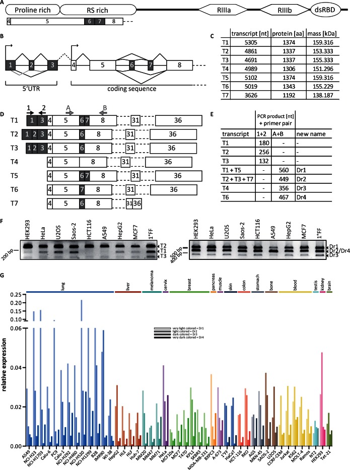
Figure 1.
Drosha transcripts and their expression in different cell lines. (A) Schematic representation of the RNase III enzyme Drosha coding region and the position of exons involved in alternative splicing. RIIIa, RNase III domain a; RIIIb, RNase III domain b; dsRBD, double-stranded RNA binding domain. (B) Schematic representation of alternative splincing. (C) Table of transcripts; nt = nucleotides; aa = amino acids; kDa = kilo Dalton. (D) Detail of the alternatively spliced region with combinations of exons both in the 5′UTR and in the N-terminal RS-rich domain in the coding sequence. T7 is additionally spliced between exon 31 and 36. Arrows indicate primer pairs for PCR amplification. (E) Expected PCR products. (F) RT-PCR results from cDNA of the indicated cell lines with primer pairs depicted in D. (G) Relative expression profile of the four identified splice variants in a set of 45 cell lines from different tissue origin, normalized to Cyclophilin A expression (N = 1 with technical duplicates).

