Figure 1.
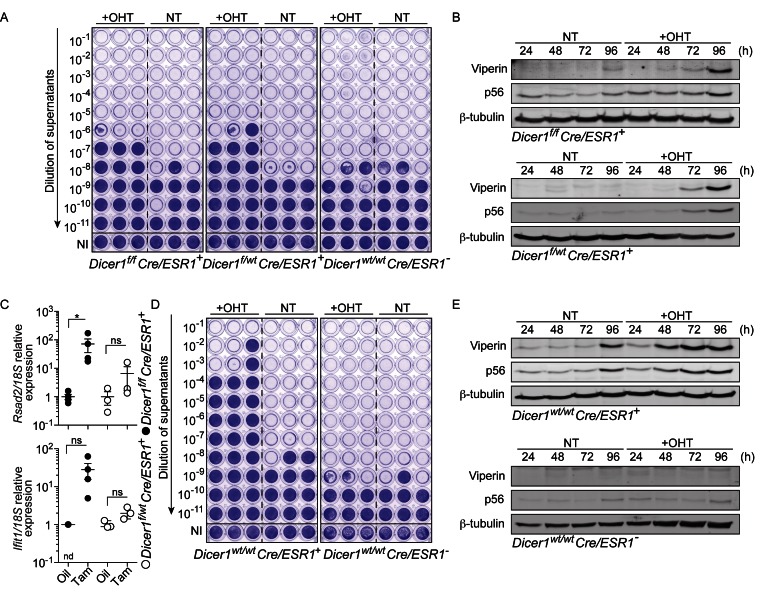
Cre-ERT-mediated activation of an antiviral response. (A) MEFs treated for 24 h with OHT were expanded for 3 days, prior to 24 h infection in biological triplicate with SFV (MOI 2). Viral titers were assayed with log10 fold dilutions on confluent Vero cells as shown. NI: not infected (uninfected cells were stained with crystal violet). (B) Time course showing the activity of 24 h OHT treatment of Dicer1f/f x Cre/ESR1+ and Dicer1f/wt Cre/ESR1 + MEFs on the levels of Viperin and p56. (A, B) Data shown are representative of a minimum of two independent experiments. NT: non-treated. (C) Peripheral blood mononuclear cells from mice injected with oil or tamoxifen for five consecutive days, and culled on day 12, were analyzed by RT-qPCR for ISG expression (each point represents one mouse; mean ± s.e.m. and unpaired Mann-Whitney U tests are shown). Data shown relative to the mean ISG/18S expression of the oil control samples. nd: non-detected in three out of four mice. (D) MEFs treated for 24 h with OHT were expanded for two days, prior to 24 h infection in biological triplicate with SFV (MOI 2) and viral titers on confluent Vero cells as in (A). (E) Time course showing the activity of 24 h OHT treatment of Dicer1wt/wt x Cre/ESR1+ and Cre/ESR1− MEFs on the levels of Viperin and p56. (C-E) Data shown are representative of three independent experiments or mice.

