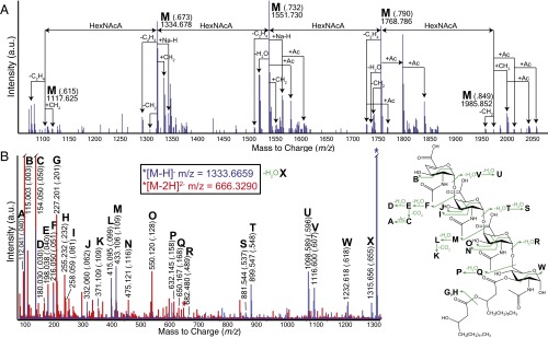
Fig. 2.
Glycolipid terminus of the Vi antigen as determined by MS. (A) Charge deconvoluted LC-electrospray ionization (ESI)-QTOF-MS spectrum in negative mode for Vi antigen termini purified from S. Typhi. All ions correspond to a di-β-hydroxyacylated HexNAc residue linked to two or more variably O-acetylated HexNAcA residues. (B) LC-ESI-QTOF-MS/MS data for the singly charged (blue) and doubly charged (red) ions corresponding to a GalNAcA3 oligosaccharide attached to a reducing terminal diacyl-HexNAc. Overlapping signals are colored purple. Fragmentations are illustrated in green.

