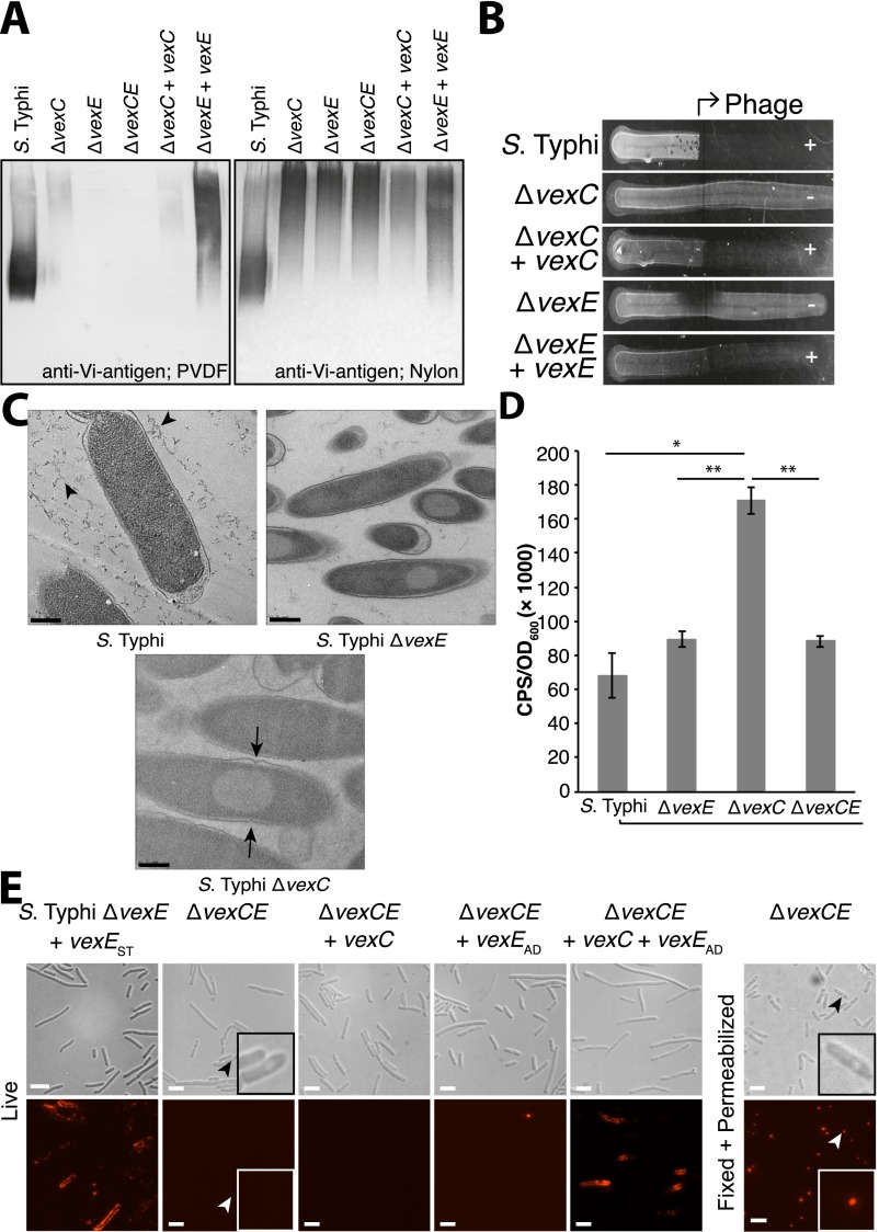
Fig. S5.
Additional phenotypic characterization of the ΔvexE mutant S. Typhi. (A) Wild-type Vi antigen bound to both hydrophobic PVDF and negatively charged nylon membranes, whereas ΔvexE mutant Vi antigen bound only to nylon. The figures are Western blots of separated proteinase K-digested whole-cell lysates, probed with anti-Vi antigen antibody. (B) S. Typhi ΔvexE is not sensitive to Vi phage II (HER#39; Félix d’Hérelle Reference Center for Bacterial Viruses, Université Laval, Québec, Canada), indicating the absence of the Vi antigen bacteriophage receptor on the cell surface. Six microliters of overnight cultures were dropped on a Petri dish in which one half of the plate had been inoculated with 7.5 × 10⁶ pfu bacteriophage (arrow). Cells are unable to grow on the phage-coated plate if they express surface-associated Vi antigen. (C) ΔvexE and ΔvexC mutants of S. Typhi accumulate cytosolic Vi antigen. Electron micrographs of thin sections of freeze-substituted S. Typhi and mutant derivatives. The hydrated Vi antigen capsule in the wild-type strain collapses during preparation (black arrowheads). The absence of export in the ΔvexC mutant and of proper acylation in the ΔvexE mutant results in electron-transparent inclusions that accumulate in the cytoplasm. Some inclusion bodies appear to interfere with cell division, because invaginations of the outer membrane are visible in the ΔvexC mutant surrounding an inclusion body (black arrows). (Scale bars, 400 nm.) (D) The Cpx cell envelope stress response is up-regulated in ΔvexC mutant but not in ΔvexE mutant S. Typhi. S. Typhi and mutant derivatives were transformed with pNLP15, which contains the spy promoter upstream of the luxCDABE cassette. Luciferase activity was monitored as luminescence counts per second and was normalized to the optical density of the cell culture. Data represent the mean ± SE of three independent experiments. Statistical differences were determined using a Students t test; significant differences are indicated: *P < 0.01; **P < 0.001. (E) Immunofluorescence microscopy of cells probed with anti-Vi antigen antibodies illustrated that the ΔvexCE mutant possessed no Vi antigen on its surface. However, the mutant accumulated intracellular Vi antigen in inclusion bodies, which became accessible to antibody in permeabilized cells. Complementation with plasmids containing vexC and vexE restored the surface expression of Vi antigen. (Scale bars, 10 µm.) Insets are enlarged to show a representative cell.

