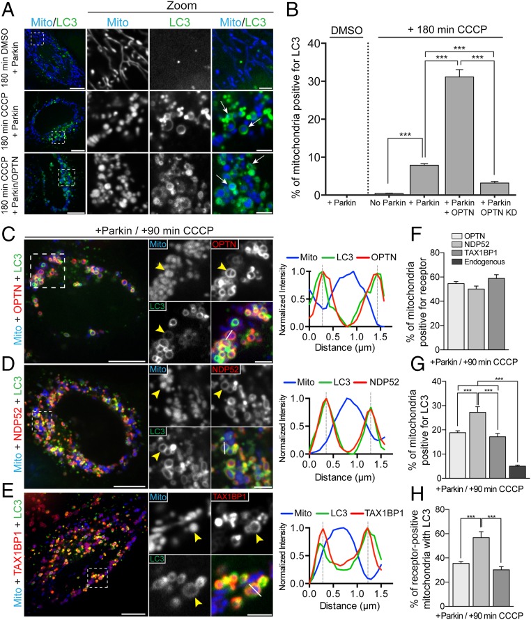
Fig. 1.
OPTN, NDP52, and TAX1BP1 are dynamically recruited to depolarized mitochondria after CCCP treatment to initiate autophagosome formation. (A) Representative confocal images showing localization of Mito-DsRed2 (blue) and GFP-LC3 (green) in Parkin-expressing HeLa cells treated with either DMSO or 20 μM CCCP. Arrows indicate mitochondria engulfed by autophagosomes. (B) Coexpression of OPTN with Parkin enhances mitophagy at 180 min post-CCCP. Depletion of endogenous OPTN significantly reduces mitophagy at this time point. KD, knockdown. (C–E) HeLa cells expressing untagged Parkin, Mito-DsRed2, and GFP-LC3, as well as Halo-OPTN (C), Halo-NDP52 (D), or Halo-TAX1BP1 (E) were treated with 20 μM CCCP for 90 min. (C) At 90 min post-CCCP, OPTN and LC3 were enriched on the outer surface of fragmented mitochondria (yellow arrowhead). Line scan analysis reveals overlapping OPTN and LC3 local maxima around peak mitochondria (Mito) fluorescence. (D) Cells expressing Halo-NDP52 exhibited stable NDP52 and LC3 recruitment to depolarized mitochondria at 90 min post-CCCP (yellow arrowheads). The corresponding line scan indicates NDP52/LC3 colocalization around the indicated mitochondrion. (E) Halo-TAX1BP1 also colocalized with LC3 around depolarized mitochondria at 90 min post-CCCP. The line scan shows the corresponding peaks of normalized LC3 and TAX1BP1 intensity. (F) OPTN, NDP52, and TAX1BP1 are robustly recruited to mitochondria after 90 min of CCCP treatment. (G and H) Expression of each receptor with Parkin enhances mitophagy at 90 min post-CCCP compared with cells expressing Parkin alone. [Scale bars: A and C–E (full size), 10 μm; A and C–E (zoom), 2.5 μm.) Data were collected from 21 to 70 cells from at least three independent experiments. Bars represent mean ± SEM. ***P < 0.001.

