Fig. 7.
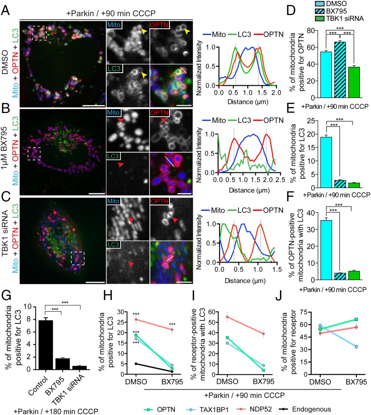
TBK1 is required for efficient autophagic engulfment of depolarized mitochondria. (A–C) HeLa cells expressing untagged Parkin, Mito-DsRed2 (blue), Halo-OPTN (red), and GFP-LC3 (green). (A) Representative confocal image of DMSO-pretreated HeLa cell at 90 min post-CCCP with the corresponding line scan. OPTN and LC3 stably localize on the surface of damaged mitochondria (yellow arrowheads). (B) Pretreatment with 1 μM BX795 significantly attenuates LC3, but not OPTN, recruitment to depolarized mitochondria (red arrowheads). The corresponding line scan indicates increased OPTN, but not LC3, fluorescence around the peak mitochondrial intensity. (C) TBK1 depletion blocks association of LC3 with fragmented mitochondria at 90 min post-CCCP (red arrowheads). The line scan indicates OPTN, but not LC3, recruitment to fragmented mitochondria. (D) Inhibition of TBK1 by BX795 increases the proportion of cellular mitochondria that recruit OPTN, whereas knockdown of TBK1 decreases OPTN-positive mitochondria. (E and F) Both TBK1 knockdown and inhibition by BX795 significantly impair autophagosome formation on OPTN-positive mitochondria at 90 min post-CCCP. (G) In Parkin-expressing cells, depletion or inhibition of TBK1 interferes with mitophagy at 180 min post-CCCP. (H) Coexpression of Parkin with either OPTN or TAX1BP1 significantly enhances mitophagy at 90 min post-CCCP in control cells, but not in cells treated with a TBK1 inhibitor. In contrast, coexpression of Parkin with NDP52 robustly enhances mitophagy independent of TBK1 activity. (I) NDP52-positive mitochondria, but not OPTN- or TAX1BP1-positive mitochondria, are efficiently engulfed by autophagosomes after TBK1 inhibition. (J) TAX1BP1 recruitment to depolarized mitochondria is diminished in cells pretreated with BX795. [Scale bars: A–C (full size), 10 μm; A–C (zoom), 2.5 μm.] Data were collected from 19 to 61 cells from at least three independent experiments. Bars represent mean ± SEM. ***P < 0.001.

