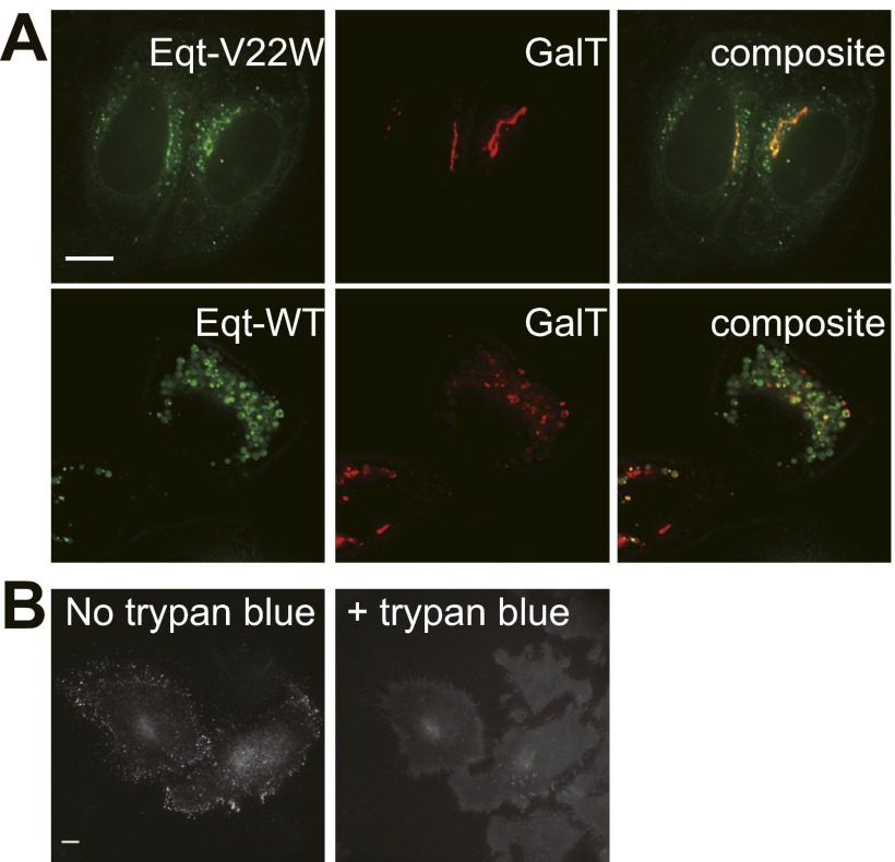
Fig. S1.
Localization of Eqt WT and V22W and trypan blue quenches fluorescence of Eqt-SM-pHlourin. (A) Localization of Eqt-V22W and native Eqt in HeLa cells. Plasmids encoding the indicated proteins were transfected into HeLa cells and visualized by deconvolution florescence microscopy at 16 h after transfection. Maximum projections of a z series are shown. (Scale bar: 10 μm.) (B, Left) Cells expressing Eqt-SM-pHlourin before the addition of trypan blue to the culture medium. (B, Right) The same cells photographed ∼30 s after addition of trypan blue (concentration) to the culture medium. Maximum projections of a z series are shown. (Scale bar: 10 μm.)

