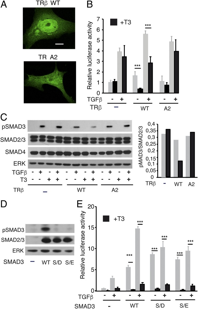
Fig. 5.
Nuclear translocation is required for T3 antagonism. (A) Representative confocal images in Mv1Lu cells transfected with WT TRβ fused to GFP and the 2A mutant in the nuclear localization signal. (Scale bar, 10 µM.) (B) Cells were transfected with an empty vector or with vectors encoding WT and 2A TRβ. Luciferase activity was measured in cells incubated with T3 for 36 h and with TGF-β for the last 5 h. (C) Western blot analysis of pSMAD3, total SMAD2/3, total SMAD4, and ERK in cells transfected with the WT and mutant receptors and treated with T3 for 36 h in the presence and absence of TGF-β for the last 30 min. (D) Western blot analysis of pSMAD3 and SMAD2/3 in Mv1Lu cells transfected with expression vectors for WT SMAD3 or with SMAD3 mutants in the SXS motif (S/E and S/D). (E) Luciferase assays in Mv1Lu cells cotransfected with TRβ and WT and mutant SMADs. After transfection, cells were incubated in the presence and absence of T3 for 24 h and/or with TGF-β for the last 5 h. Luciferase results (mean ± SD) in the different panels are expressed relative to the values obtained in untreated cells transfected with empty vectors. Statistically significant differences between cells treated with and without T3 are indicated.

