Fig. 8.
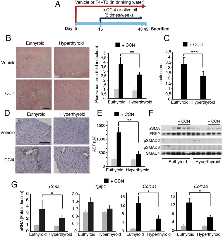
Administration of thyroid hormones attenuates liver fibrosis induced by CCl4 treatment in mice. (A) Scheme of the treatments (vehicle, n = 4; CCl4, n = 6). (B) Representative Picrosirius red staining of livers of euthyroid and hyperthyroid mice after chronic treatment with vehicle or CCl4. (Scale bar, 400 µm.) Fibrosis was quantified from the stained area. (Right) Results (mean ± SEM) are shown. (C) Fibrosis grading with the Ishak score obtained with the same samples. (D) Immunohistochemistry of Collagen1 expression. (Scale bar, 200 µm.) (E) Serum AST activity (mean ± SEM). (F) Western blot of α-SMA, pSMAD2, and pSMAD3 in livers after chronic CCl4 treatment. ERK2 and SMAD4 were used as loading controls. (G) Relative mRNA levels of the fibrogenic genes α-Sma, Tgfβ1, Col1a1, and Col1a2. Results (mean ± SEM) are expressed relative to the values obtained in control euthyroid mice treated with vehicle. Statistically significant ANOVA posttest differences (Bonferroni) between CCl4-treated euthyroid and hyperthyroid groups are shown.

