Fig. 9.
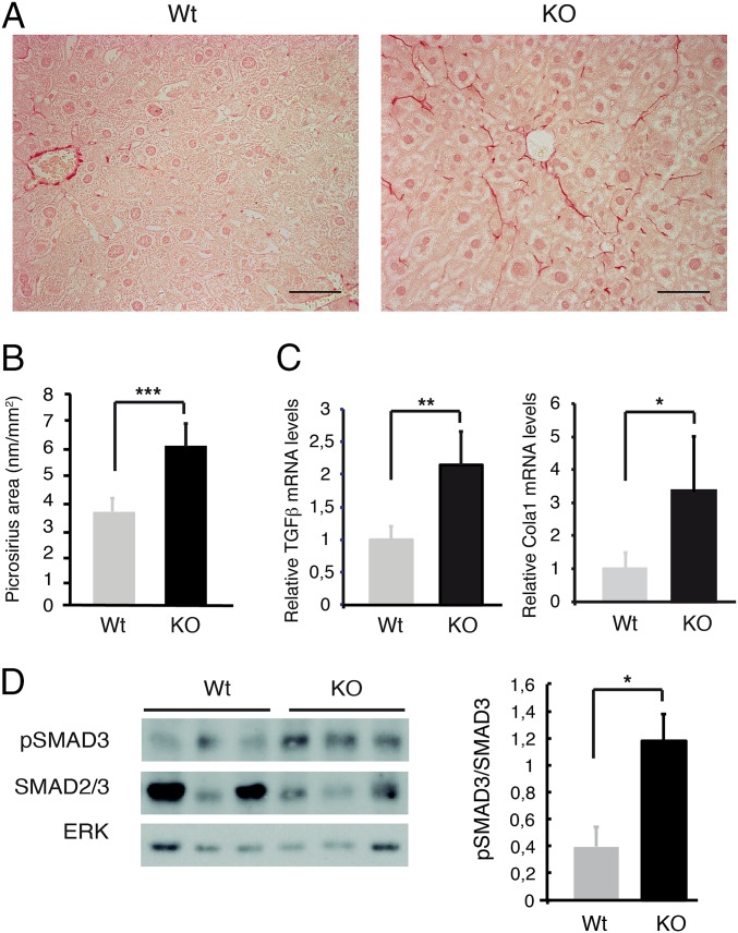
Genetic deletion of TRs causes spontaneous liver fibrosis in aged mice. (A) Picrosirius red staining in representative liver sections of WT mice (n = 9) and KO mice (n = 5) lacking the thyroid hormone binding isoforms TRα1 and TRβ. (Scale bar, 50 µm.) (B) Quantification (mean ± SEM) of the Picrosirius red stained area. (C) Relative levels of Tgfβ1 and Col1a1 mRNA in both groups. (D) Western blot of pSMAD3, total SMAD2/3, and ERK in livers from WT and KO mice. (Right) pSMAD3/SMAD2,3 ratio (mean ± SEM) obtained in four WT and six KO mice. Statistically significant unpaired two-tail Student´s t test differences between WT and KO groups are shown.

