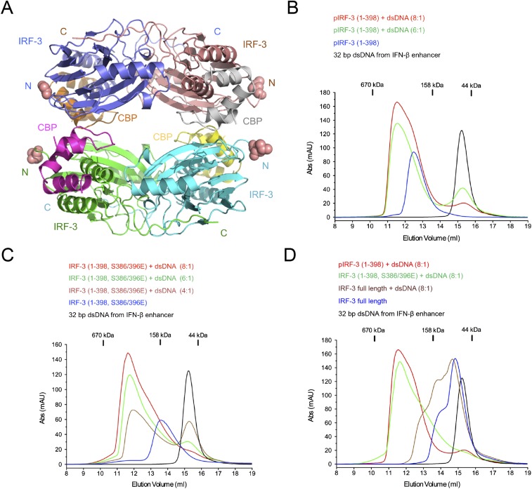
Fig. S6.
Structure of the IRF-3/CBP tetramer in the ASU and binding studies of the pIRF-3 (residues 1–398)/CBP complex, IRF-3 (residues 1–398, S386/396E)/CBP complex, and unphosphorylated full-length IRF-3 with a 32-bp dsDNA derived from the IFN-β enhancer. (A) Crystallographic tetramer of IRF-3 (residues 189–398, S386/396E) in complex with CBP. (B) Gel-filtration chromatography DNA-binding studies of pIRF-3 (residues 1–398) in complex with CBP. The sequence of the DNA used in this study is 5′-CATAGGAAAACTGAAAGGGAGAAGTGAAAGTG-3′. (C) Gel-filtration chromatography DNA-binding studies of the IRF-3 (residues 1–398, S386/396E) mutant in complex with CBP. (D) Gel-filtration chromatography DNA-binding study of unphosphorylated full-length IRF-3.

