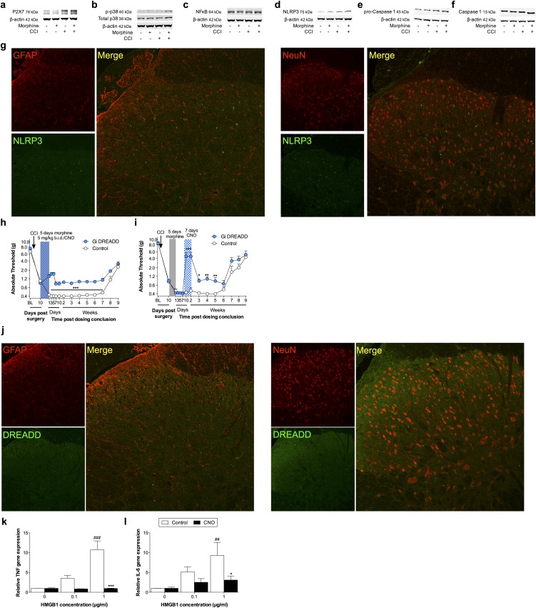
Fig. S4.
(A–F) Ipsilateral lumbar dorsal spinal cords were collected from F344 rats that had undergone sham or CCI surgery, 5 wk after morphine/saline administration, and respective levels of P2X7R (A), phospho-p38/total p38 ratio (B), NF-κB (p65 subunit) (C), NLRP3 (D), procaspase-1 (E), and caspase-1 (F) quantified. Representative blots presented. (G) Ipsilateral lumbar dorsal spinal cords were collected from F344 rats that had undergone sham or CCI surgery, 5 wk after morphine/saline administration. NLRP3, GFAP, and NeuN immunofluorescence. (H and I) SD rats were transfected with intrathecal inhibitory Gi or control DREADDs, and morphine (5 d; shaded area) was administered 10 d after CCI, and absolute thresholds for mechanical allodynia quantified in F344 rats. CNO (blue hatched bar) was coadministered with morphine (5 d) (H) or 1 wk after morphine conclusion (7 d) (I), and absolute thresholds for mechanical allodynia quantified. (J) Ipsilateral lumbar dorsal spinal cords were collected from F344 rats, 21 d after transfection with control DREADDs. DREADD, GFAP, and NeuN immunofluorescence. (K and L) TNF (K) and IL-6 (L) gene expression in BV-2 cells expressing the Gi DREADD after 4 h incubation with a concentration range of HMGB1, and 0 μM (control) or 50 μM CNO. *P < 0.05; **P < 0.01; ***P < 0.001 (relative to control); ##P < 0.01: ###P < 0.001 (relative to 0 μg). Data are presented as mean ± SEM; n = 6 or 7 per group.

