
Figure 2. Melatonin expression evaluated by immunofluorescence confocal microscopy (400x) in the pineal gland (A) and thymus (B) of elderly people.
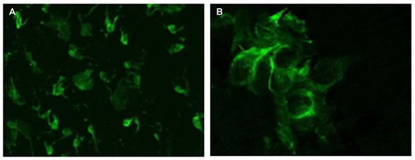
Samples were fixed in 4% neutral buffered formalin (pH 7.2) for 15 minutes and incubated with primary monoclonal mouse antibodies to melatonin (Mubro Products B.V., 1:50). After washout in phosphate buffer, samples were incubated with a second antibody, rabbit antimouse FITC-conjugated Ig (Dako, 1:100). After washout in phosphate buffer, a medium containing 90% glycerin, 0.02M Tris-HCl (pH 8.0), 0.8% NaN3, and 2% 1.4-di-azabicyclo-(2,2,2)-octan (Sigma-Aldrich) was added to the cells. The preparations were examined under a Leica TCS SP5 confocal microscope using an MRC-1024 system equipped with LaserSharp 5.0 software (Bio-Rad) for confocal image analysis.