
Figure 4. CD5 expression evaluated by immunohistochemistry (200x) in the pineal gland (A) and thymus (B) of elderly people.
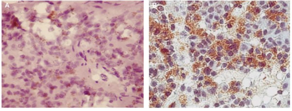
Samples of pineal gland and thymus were fixed in formalin at standard pH, washed with ethanol solutions and fixed using a paraffin-embedding standard technique. Slices with a thickness 3-5 μm were made using a Leica 540M microtome and placed on slides with poly-L-lysine. For histological analysis the samples were stained using the standard hematoxylin-eosin staining technique. The identification of CD5 (1:30, Novocastra) was carried out with an immunohistochemical method using primary mouse monoclonal antibodies, and secondary antibodies, i.e. biotinylated anti-mouse immunoglobulins (Novocastra). Visualization of the reaction was carried out using the avidin-peroxidase complex (ABC-kit) and diaminobenzidine.