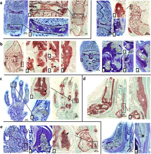
Figure 2. Aberrant cartilage and ligament mineralization in asj-2J mice at 12 weeks of age.

Toluidine Blue O staining (blue & purple) and Alizarin red staining (red & green) reveal extensive cartilage and ligament mineralization in the intervertebral disc, knee, foot, ankle and shoulder of asj-2J mice, marked by arrow or asterisk. a. Coronal sections of spine show mineralization in intervertebral discs and posterior longitudinal ligament (1). b. Coronal and sagittal sections of the knee show mineralized in patellar tendons (2), posterior/anterior cruciate ligaments (3), meniscus (5) and lateral/medial collateral ligaments (4,6). c. Coronal sections of the foot show the hypertrophied mineralized joints (7). d. Sagittal plane of the ankle shows the Achilles tendon mineralization (8). e. Coronal sections of shoulder show supraspinatus tendon mineralization (9).
