
Figure 7. Blocking of sirt1 activity using siRNA and sirtinol inhibited the activation of autophagy flux by melatonin.
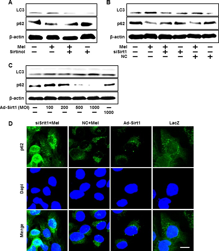
HaCaT keratinocytes were incubated with melatonin in the presence of sirtinol for 24 hr. Total keratinocyte extracts were prepared and analyzed by Western blot for LC3-II and p62 protein levels A. Sirt1 small interfering RNA (siSirt1) or negative control siRNA (NC siRNA) transfected HaCaT keratinocytes were incubated with 10 μM of melatonin. Western blot for LC3-II and p62 proteins was analyzed from HaCaT keratinocytes B. HaCaT keratinocyte was transfected by overexpressing adenovirus (Ad-Sirt1) or lacZ-bearing adenovirus (Ad-lacZ). Western blot for LC3-II and p62 proteins were analyzed from HaCaT keratinocytes C. β-actin was used as loading control. The cells were immunostained with p62 antibody (green) and observed in the fluorescent view Scale bar: 10 μm D.