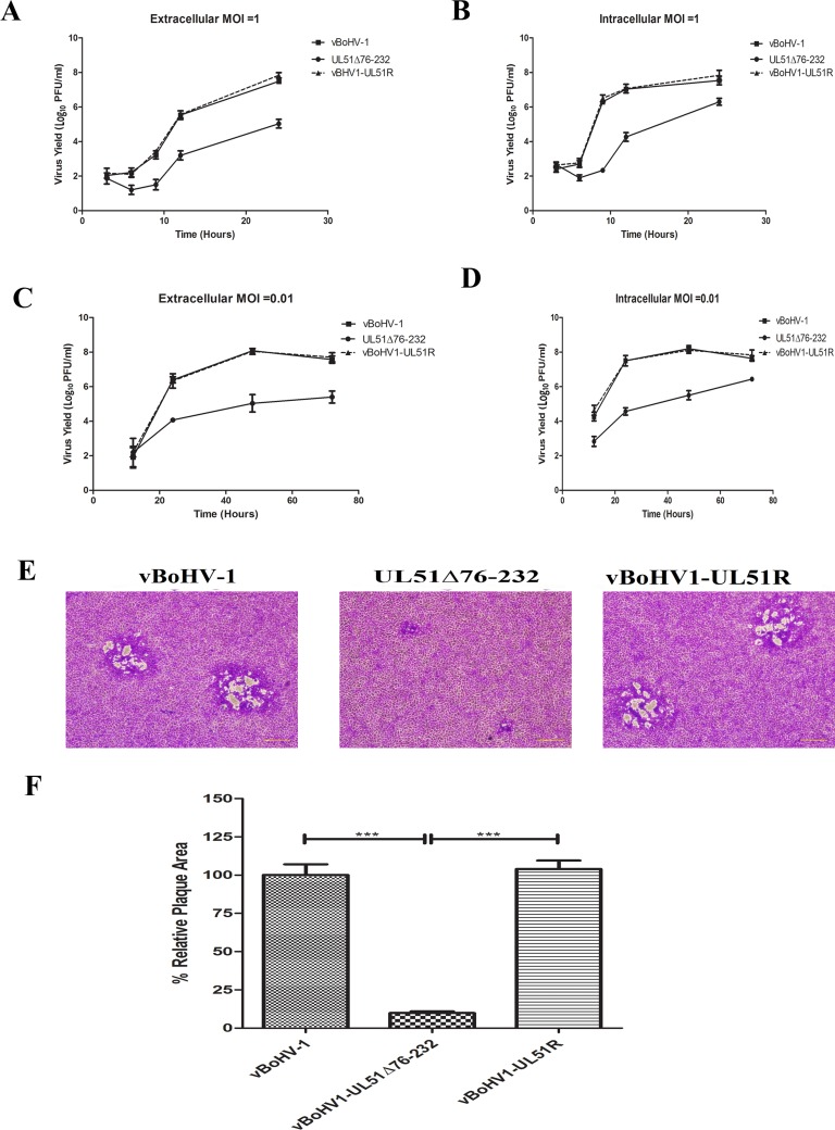
Figure 4. Growth kinetics of the UL51 mutant virus in MDBK cells.
Growth of the wild-type (■), UL51Δ76-232 mutant (●) and UL51 revertant (▲) viruses. For single-step growth kinetics, MDBK cells were infected with an MOI of 1. At 3, 6, 9, 12, and 24 hpi, extracellular A. and intracellular B. virus particles were collected, and virus yields were determined by titration on MDBK cells. Each data point represents the mean ± standard deviation of three independent experiments. For multi-step growth kinetics, MDBK cells were infected with the indicated viruses at an MOI 0.01. Extracellular C. and intracellular D. virus particles were sampled at 12, 24, 48, and 72 hpi, and titrated on MDBK cells. Each data point represents the mean ± standard deviation of three independent experiments. E. Plaque morphology and plaque size of the vBoHV-1, UL51Δ76-232, and vBoHV1-UL51R viruses. MDBK cells were infected with 200 PFU per well of the indicated viruses, and overlaid with methylcellulose. After 48 h, the cells were fixed and stained with crystal violet. F. The plaque area was analyzed with an Olympus IX70® Microscope. The mean percentages of the plaques and standard errors were determined by counting 50 random plaques of each virus, and the significance level was calculated using a t-test (***, P < 0.0001).

