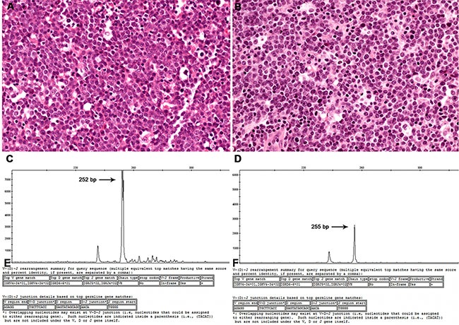
Figure 1. Representative case (#4).

Histopathology sections of a lymph node diagnosed as a DLBCL in both 2002 (A) and 2004 (B). Using GeneMapper analysis, a peak at 252 bp was seen in the primary DLBCL (C), whereas a peak at 255 bp appeared in the relapsed DLBCL (D). In sequencing analysis, both tumors showed identical VH, DH, and JH gene segments and junctions (E and F).
