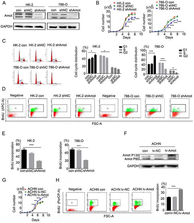
Figure 3. Amot promotes proliferation of renal epithelial and RCC cells.

A. Western blot analysis of Amot silencing in HK-2 and 786-O cells. B. Knockdown of Amot inhibits HK-2 and 786-O cell proliferation. The shAmot group of cells was compared with control group or shNC group of cells. C. Knockdown of Amot induces cell cycle arrest at G1 phase in HK-2 and 786-O cells. D, E. Knockdown of Amot inhibits BrdU incorporation in HK-2 and 786-O cells using APC-anti-BrdU. Negative: The cells were treated with PBS in place of Anti-BrdU antibody as the negative control. F. Western blot analysis of up-regulated Amot p130 expression in ACHN/lv-Amot cells. G. Enhanced Amot expression promotes ACHN cell proliferation. The lv-Amot group of cells was compared with the control group or lv-NC group of cells. H. Up-regulated Amot expression enhances BrdU incorporation in ACHN/lv-Amot cells using PerCP-Anti-BrdU. Negative: The cells were treated with PBS in place of Anti-BrdU antibody as the negative control. Con: Unmanipulated cells; NC (Negative control): the cells infected with control lentivirus; shAmot (Amot knockdown); The cells infected with shAmot lentivirus; lv-Amot (Up-regulated Amot expression): The cells infected with lv-Amot lentivirus. Data are representative images and expressed as the mean ± SD of each group of cells from three separate experiments. *P < 0.05; **P < 0.01; ***P < 0.001.
