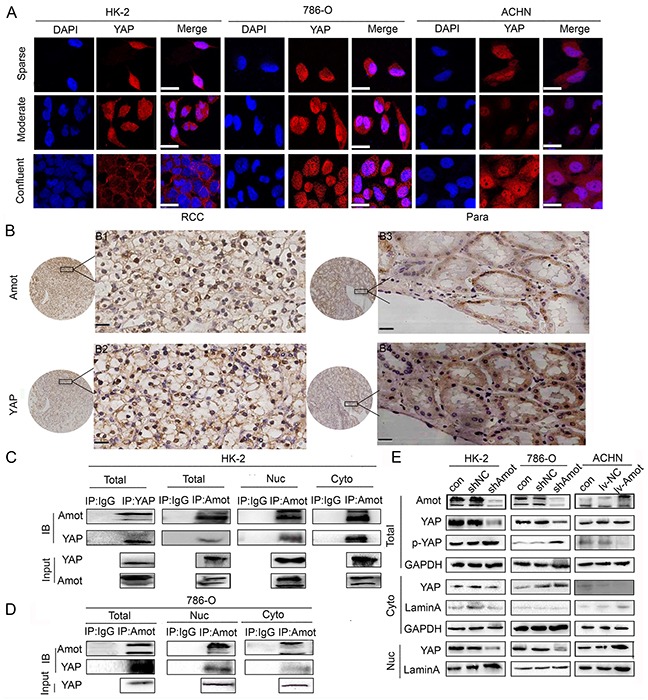
Figure 5. Amot is crucial for retaining the nuclear YAP.

A. Immunofluorescence of Amot in HK-2,786-O and ACHN cells cultured at sparse, moderate and confluent density. Sparse: 30% density, moderate: 60%-80% density, confluent: 100% density; Scale bar = 25μm B. Immunohistochemistry of Amot and YAP expression in human RCC and paracancerous tissues. Para: paracancerous tissues; B1, B3: anti-Amot; B2, B4: anti-YAP; Scale = 33μm. C. Immunoprecipitation and Western blotting analysis of endogenous Amot-YAP complexes in HK-2 cells (IB). Western blot analysis of Amot and YAP in cell lysates from HK-2 cells (Input). Rabbit IgG from unvaccinated animals was used as the control. D. Immunoprecipitation and Western blotting analysis of endogenous Amot-YAP complexes in 786-O cells (IB). Western blot analysis of Amot and YAP in cell lysates from 786-O cells (Input). E. Western blot analysis of Amot, total YAP, phosphorylated YAP, GAPDH, and Lamin A in cytoplasmic and nuclear fractions of cells. GAPDH was used as an internal control. Lamin A was used as a nuclear marker.
