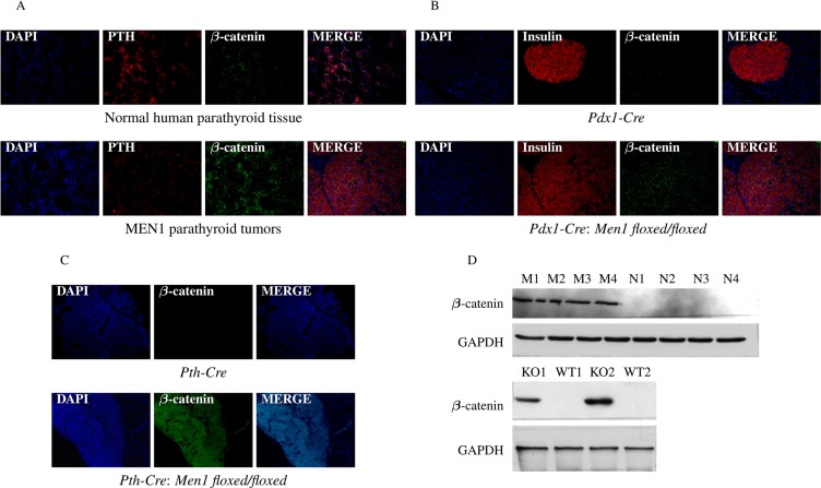
Figure 9. Hypermethylation of Sox genes results in increased β-catenin expression.
The presence of β-catenin was significantly increased in parathyroid tissues from MEN1 patients (A), mouse pancreatic endocrine tumors (B), and mouse parathyroid tumors (C) from Men1 KO mice compared to normal endocrine tissues from humans and control mice by IF staining. PTH and insulin were stained red by anti-PTH antibody and anti-insulin antibody, respectively (Alexa Fluor 647); β-catenin was stained green by anti-β-catenin antibody (Alexa Fluor 488); and nuclei were stained blue with DAPI. Images were taken with the 20X objective. Increased β-catenin expression in parathyroid tumors from MEN1 patients and pancreatic endocrine tumors from Men1 KO mice was further confirmed by western blot assay (D).

