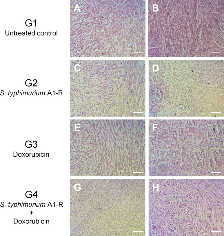
Figure 3. Histological response.
Untreated tumors (G1) were comprised of spindle-shaped viable sarcomatous cells without necrosis or inflammatory change A. B. Tumors treated with S. typhimurium A1-R (G2) had components of inflammation (C. Grade IIB), or necrosis (D. Grade IIA). One of the tumors treated with doxorubicin (DOX) (G3) showed no necrosis (E. Grade I), while another tumor showed complete tumor necrosis (F. Grade IV). Tumors treated with the combination of S. typhimurium A1-R and DOX were destroyed and replaced with inflammatory-type cells (G. H. Grade III). Hematoxylin and eosin staining. Scale bars: 100 μm.

