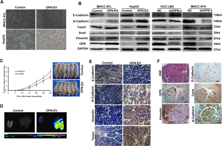
Figure 1. OPN promotes EMT in HCC cells.
(A) Representative pictures show the morphological change after overexpression of OPN in HCC cells. (B) Expression of EMT-related biomarkers were detected in MHCC-97L and HepG2 cells with up-regulation of OPN (left two panels) or HCC-LM3 and MHCC-97H cells with OPN knockdown (right two panels). (C) Both in vivo tumor growth rates (left panel) and tumor sizes (right panel) in mice models subcutaneously implanted with HepG2-OPN cells were much larger than that of those controls (P < 0.05). (D) The visual and fluorescent images demonstrated obviously stronger fluorescent signals in both liver and lung of nude mice models subcutaneously implanted with HepG2-OPN cells (right), while no obvious GFP signal was detected in liver or lungs of the controls (left). (E) Immunohistochemical staining for the expressions of EMT-related markers in subcutaneous xenografts tumor tissues from nude mice models of OPN-upregulated HepG2 cells and the controls (Magnification × 400. Bar = 50 μm). (F) Representative HCC cases in tissue slides (serial sections) were analyzed by H & E and immunohistochemical staining for OPN and EMT-related markers. (Magnification × 100. Bar = 200 μm).

