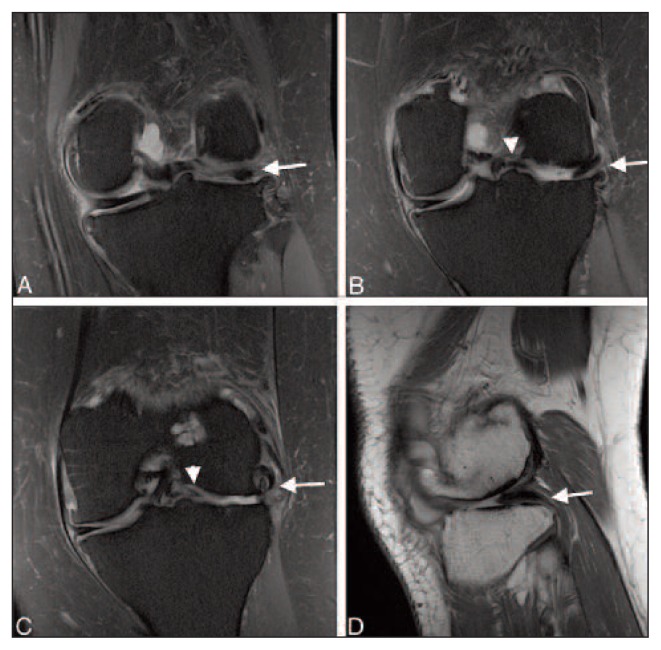
Figure 2.
Sequential coronal proton density with fat saturation images, from posterior to anterior (a–c), of a 46-year-old female with a medially displaced popliteus tendon (white arrows) residing within the lateral intercondylar space and a medially displaced lateral meniscus (white arrowheads). Sagittal proton density (d) weighted image also shows the popliteus tendon in the lateral intercondylar space (white arrow).

