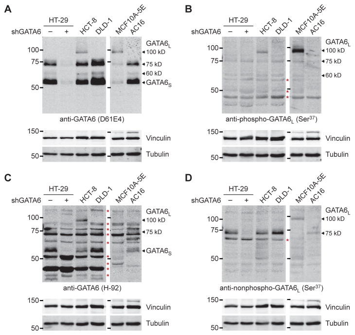
Fig. 9. Diversity of GATA6L forms across different cell lineages.
(A to D) Arrows indicate the GATA6 forms confirmed earlier by knockdown or observed with multiple antibodies. Red asterisks indicate nonspecific bands. The MCF10A-5E and AC16 samples are on an immunoblot; HT-29, HCT-8, and DLD-1 are on another blot. The blots have been scaled to match. Data are representative of n ≥ 3 independent experiments.

