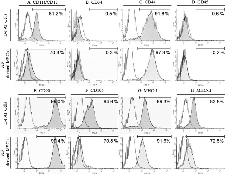
Fig. 4.
Results of flow cytometry using immunological markers on DFAT cells and AT-MSCs. A strong shift in MFI was detected with antibodies against CD44 (C), CD90 (E), and MHC class I (G); a positive signal with antibodies against CD11a/18 (A), CD105 (F), and MHC class II (H) partially overlapped with the negative control; and no positive signal was detected with antibodies against CD34 (B) and CD45 (D). The dotted line represents the negative control. The horizontal line in individual histograms indicates the population (%) of positive cells. DFAT, dedifferentiated fat; AT-MSCs, adipose tissue-derived mesenchymal stem cells; MFI, mean fluorescence intensity; MHC, major histocompatibility complex.

