Figure 3.
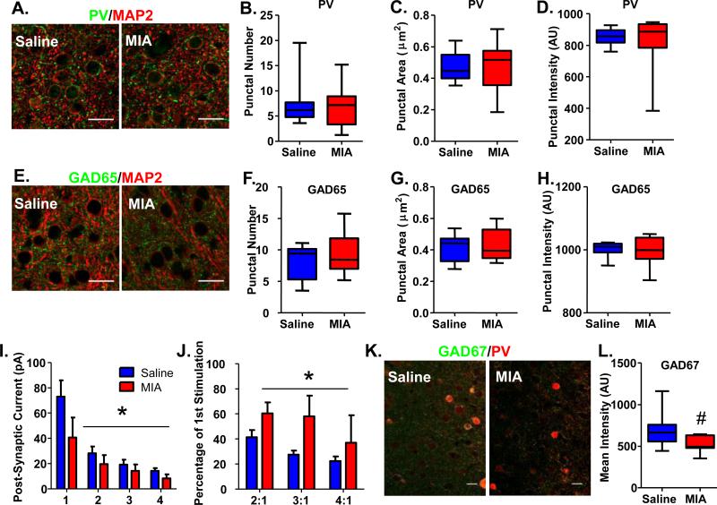
GAD65 and PV perisomatic puncta are not altered, but presynaptic release probability and GAD67 expression is decreased in PV cells in the mPFC of adult MIA offspring. (A) Examples of PV/MAP2 staining of the mPFC of Saline and MIA offspring. (B) The number, (C) area and (D) intensity of PV-expressing perisomatic puncta surrounding pyramidal cells in the mPFC are unaltered in adult MIA offspring. (E) Examples of GAD65/MAP2 staining of the mPFC of Saline and MIA offspring. (F) The number, (G) area and (H) intensity of GAD65-expressing perisomatic puncta surrounding pyramidal cells in the mPFC are also unchanged in adult MIA offspring. (I) Four 5 ms pulses of blue light were delivered at 20 Hz to slices of mPFC from adult MIA or Saline offspring expressing ChR2 in PV cells. The amplitudes of the post-synaptic currents evoked in pyramidal cells by light stimulation were significantly smaller in MIA offspring. (J) The ratio of the amplitudes of the post-synaptic currents evoked in pyramidal cells by the 2nd, 3rd and 4th stimulation with light compared with the 1st stimulation were significantly increased in MIA offspring. (K) Examples of GAD67/PV staining of the mPFC of Saline and MIA offspring. (L) The mean intensity of GAD67 staining within PV cells exhibited a strong trend towards being decreased in MIA offspring. Scale bar = 20 μm. *p<0.05. #p=0.07. (See also Supplementary Figure S3 and S4).

