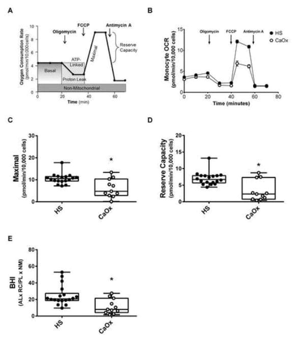
Figure 1.
Mitochondrial function in monocytes from calcium oxalate (CaOx) stone formers and healthy subjects (HS). (A) Illustration of the mitochondrial assay used to measure oxygen consumption rate (OCR) over time using the mitochondrial stress test. (B) OCR profiles of monocytes from healthy subjects (black circle) and CaOx stone formers (white circle) using the mitochondrial stress test. Indices of mitochondrial function—(C) maximal respiration and (D) reserve capacity were calculated from all study participants based on their respective bioenergetic profile. (E) The BHI was calculated using the equation: (ATP-linked OCR x Reserve Capacity)/(Proton Leak x Non-mitochondrial OCR). The indices are represented in a box plot with lower 25th percentile, median, upper 75th percentile, and whiskers drawn at 1.5 × interquartile range. Results are n=18 healthy subjects and n=11 CaOx stone formers. *p<0.05 compared to healthy subject monocytes.

