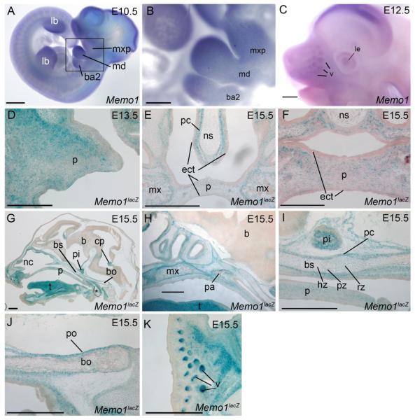
Figure 4. Embryonic expression of Memo1 during craniofacial development.
(A-C) Lateral views of embryos processed by whole-mount in situ hybridization using an anti-sense Memo1 riboprobe at E10.5 (A, B) and E12.5 (C). Note, (B) is boxed region in (A) shown at higher magnification. (D-K) b-galactosidase staining (blue) of frozen Memo1neo/+ sections at the time points indicated in either a frontal (D-F) or sagittal (G-K) plane, counterstained with nuclear fast red. A single palatal shelf is shown at E13.5, prior to fusion (D), or both shelves at E15.5 subsequent to secondary palatal fusion (E, F). Note, E is slightly more posterior than F. Additional images from E15.5 show a midsagittal plane of the entire embryonic head (G) or detailed images of the secondary palate and anterior cranial base (H), mid-cranial base (I), posterior cranial base (J), and snout (K). Orientation for (G-K) is rostral left, caudal right. Abbreviations: b, brain; ba2, branchial arch 2; bs, basisphenoid; bo, basioccipital; cp, choroid plexus; ect, ectoderm; hz, hypertrophic zone; lb, limb bud; le, lens; md, mandibular prominence; mx, maxilla; mxp, maxillary prominence; nc, nasal cavity; ns, nasal septum; p, palate; pa, palatine bone; pc, perichondrium; pi, pituitary gland; po, periosteum; pz, proliferative zone; rz, resting zone; t, tongue; v, vibrissae. Note, asterisk in G. highlights air bubble from processing. Scale bars: A, C, E, G-K: 500uM; B: 125uM; D, F: 200uM

