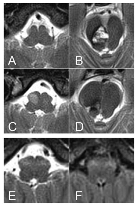
Figure 1. MR images of example cases who had unilateral and bilateral hypertrophic olivary degeneration (HOD).
(A–D): unilateral HOD. (E, F): bilateral HOD. On a day before surgery, there was no abnormal signals in the medulla (A), but a large cavernous malformation with evolving blood products was evident in right pontomesencephalic junction (B). Four months after resection of the cavernous malformation, a newly developed hyperintense lesion was observed in right ION (C), while there were only postoperative changes in pontomesencephalic junction (D). At age of 54 years, symmetrical hyperintense lesions were detected in bilateral ION (E, F). (A–E, T2-weighted images; F, fluid-attenuated inversion recovery image)

