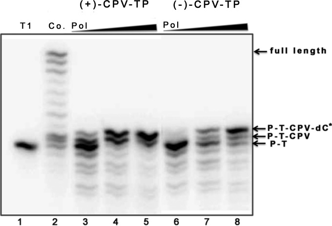
FIG 4.
DNA extension by HCMV DNA polymerase following CPV incorporation. Radiolabeled primer template T1 (Fig. 2A) was incubated with either dGTP, (+)-CPV-TP, or (−)-CPV-TP, dATP, dCTP, and dTTP, and various concentrations of HCMV Pol (7.2 nM, lanes 2, 3, and 6; 36 nM, lanes 4 and 7; 72 nM, lanes 5 and 8; increasing polymerase concentrations indicated as wedges above the panel) in the presence of UL44, and the products were analyzed by polyacrylamide gel electrophoresis and autoradiography. Lane 1, untreated radiolabeled T1; lane 2, DNA extension by HCMV Pol and UL44 in the presence of all four deoxynucleoside triphosphates without CPV-TP (Co.); lanes 3 to 5, DNA extension in the presence of (+)-CPV-TP; lanes 6 to 8, DNA extension in the presence of (−)-CPV-TP. The arrows at the right of the panel indicate the major species observed. P-T, unmodified primer template T1; P-T-CPV, T1 with either (+)-CPV-TP or (−)-CPV-TP added; P-T-CPV-dC*, T1 with either (+)-CPV-TP or (−)-CPV-TP and the next nucleotide added, the asterisk indicating that it has not been established that this residue is dC; full length, the largest product observed in lane 2.

