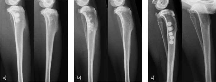
FIG 2.
X rays of tibia bones after implantation of bone substitute materials. (a) Gentamicin-containing beads (CaSO4-G [Herafill]); (b) calcium sulfate beads carrying vancomycin (CaSO4-V); (c) commercially available tobramycin-loaded calcium sulfate beads (Osteoset). In each panel, the image on the left was taken immediately after implantation and that on the right after 12 weeks in vivo.

