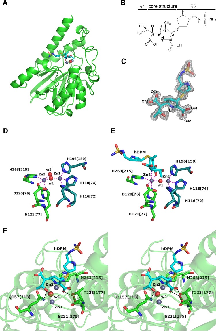
FIG 1.
(A) Ribbon representation of SMB-1 (green) bound to hDPM (cyan sticks). Zinc ions and the water molecule are depicted as gray and red spheres, respectively. (B) Chemical structure of hDPM. (C) 2|Fo|-|Fc| electron density map contoured at 1.0 σ (gray mesh). hDPM is shown as cyan (carbon), ocher (sulfur), red (oxygen), and blue (nitrogen) sticks. (D) Coordination of zinc ions in native SMB-1. Zinc ions and water molecules are depicted as gray and red spheres, respectively. Amino acids coordinating Zn-1 are Zn-2 are illustrated as dark green and green sticks, and coordination bonds are shown as magenta dashed lines. The image was adapted from the structure with PDB accession no. 3VPE. (E) Coordination of zinc ions in hDPM-bound SMB-1. hDPM is illustrated as in panel C, while other elements are rendered as in panel D. (F) Interactions other than coordination bonds between SMB-1 and hDPM. Amino acids are shown as green (carbon), red (oxygen), and blue (nitrogen) sticks, while hDPM is illustrated as in panel C. Water molecule w1 and zinc ions Zn-1 and Zn-2 are shown as in panel A. Hydrogen bonds are shown as black dashed lines.

