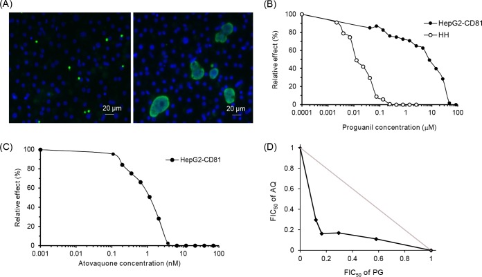
FIG 1.
Inhibitory effect of atovaquone and proguanil on the development of liver stage rodent Plasmodium infection. P. yoelii sporozoites were added to HepG2-CD81 cell monolayers together with different drug concentrations. The drug-containing cell culture media were replaced 2 and 24 h after infection, and the development of liver stage parasites was stopped 48 h after infection by fixation with methanol. Liver stage parasites were stained with a polyclonal antibody specific for Plasmodium HSP70 (green), while host and parasite nuclei were stained with DAPI (blue). The numbers of arrested and nonarrested parasites were evaluated by microscopic examination. (A) Representative image of P. yoelii parasites 48 h after infection. Small arrested parasitic forms (<7 μM) with a single nucleus are shown in the left panel, and fully developed schizonts are displayed in the right panel. (B) Dose-response curves for proguanil against P. yoelii in HepG2-CD81 and primary human hepatocytes (HH). Data are representative of those from three independent assays. (C) Dose-response curve for atovaquone against P. yoelii in HepG2-CD81 cells. Data are representative of those from three independent assays. (D) Isobologram showing the interaction between atovaquone and proguanil against P. yoelii infecting HepG2-CD81 cells. Drug interactions are represented by the normalized FIC50s for atovaquone and proguanil plotted against each other. Data are representative of those from three independent assays. The gray line is the line of additivity.

