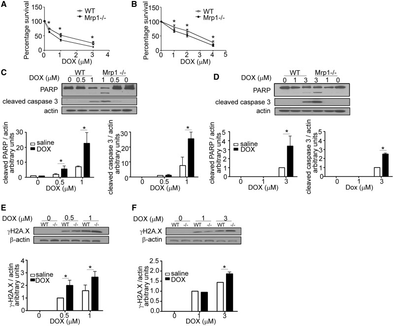
FIG. 2.
Effects of DOX on cell viability and DNA damage in wild type (WT) and Mrp1−/− cells. CM (A) and CF (B) were cultured on a 96-well plate for 48 h before treatment with varying concentrations of DOX for 3 h, followed by incubation in fresh medium. Tetrazolium reduction was measured 48 h after DOX removal. Each point represents the mean ± SD from 3 independent experiments (*P < .05 by Student’s t test). The greater increase of cleaved poly (ADP-ribose) polymerase 1 (PARP), cleaved caspase-3 and γH2A.X in CM (C and E) and CF (D and F) derived from Mrp1−/− mice was detected by immunoblot assays 24 h after 3 h of DOX treatment. The blots are representative of 1 of 3 independent experiments. Each bar represents the mean ± SD. (*P < .05 by Newman-Keuls multiple comparison test after 1-way ANOVA).

