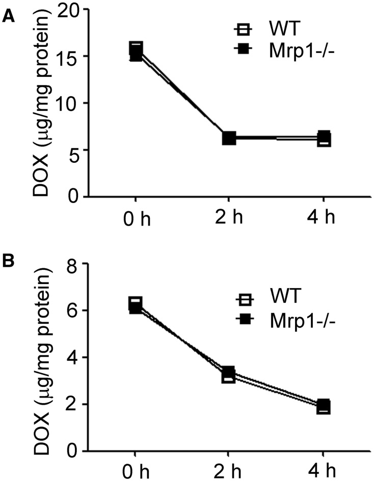
FIG. 3.
Accumulation of DOX in WT and Mrp1−/− CM and CF. Cells were treated with 30 µM DOX for 1 h, cells washed 3 times with PBS and incubated with fresh DMEM medium for the indicated times and DOX fluorescence by spectrofluorometry. The value of DOX accumulation within the cells was calculated according to the standard curve. Each point represents the mean ± SD (n = 3).

