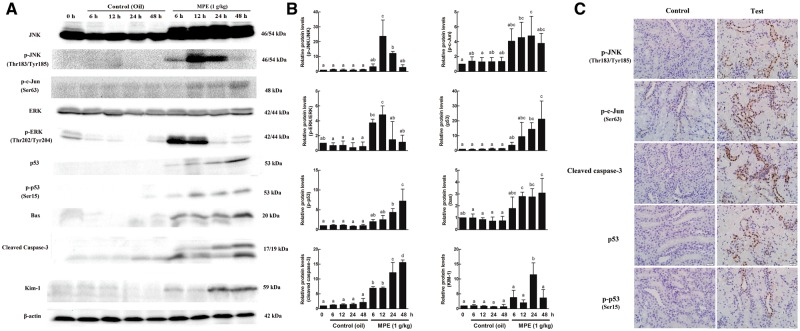
FIG. 5.
Effects of 3-MCPD palmitate on the protein levels in the kidney of Sprague Dawley rats using Western blot and immunohistochemistry (IHC) staining. A, Representative Western blot. Western blot was performed using antibodies against JNK, p-JNK, p-c-Jun, ERK, p-ERK, p53, p-p53, bax, cleaved caspase-3, and Kim-1. β-actin was used as a loading control. B, Quantitated Western blot data using Image Lab software. Western blots from 3 animals were quantitated as described in Materials and Methods. Different letters represent significant differences at P < .05. C, Representative IHC of kidney cortex tubular cells for localization of p-JNK, p-c-Jun, cleaved caspase-3, p53, and p-p53 (×200 magnification).

