Figure 5.
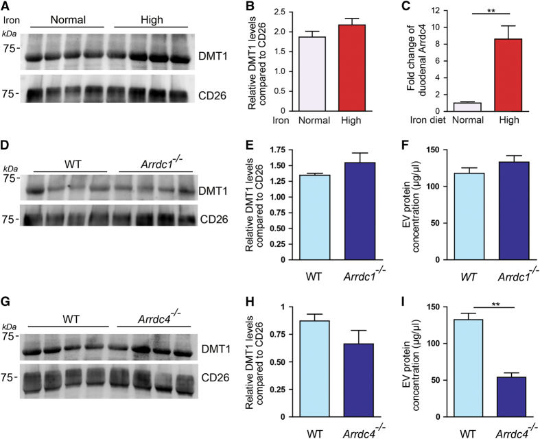
Endogenous DMT1 is released in EVs from mouse gut explants. (A) Endogenous DMT1 is released in EVs by mouse gut explants under normal and high iron conditions. CD26 is used as a loading control. (B) Densitometry quantification of DMT1 release in EVs from the gut normalized against CD26 shows a trend for increase in the amount of DMT1 released under high iron conditions. Data are mean±s.e.m.; n=4 animals per group. (C) Quantitative PCR (Q-PCR) shows a ~8.5-fold increase in the expression of Arrdc4 in the duodenum of mice fed a high iron diet compared with normal iron diet. The TATA box binding protein was used as the reference gene. Data are mean±s.e.m., n=3–4, **P<0.01. (D) DMT1 is released in gut EVs from WT and Arrdc1 −/− mice. (E) Densitometry quantification of DMT1 release in EVs from the gut normalized against CD26 shows that the levels of DMT1 released in gut EVs is not changed in Arrdc1 −/− compared with WT mice. (F) The protein concentration of EVs released from Arrdc1 −/− gut EVs is the same as from WT gut EVs. Data are mean±s.e.m.; n=4 animals per group. (G) DMT1 is released in gut EVs from WT and Arrdc4 −/− mice. (H) The levels of DMT1 released in gut EVs is not changed in Arrdc4 −/− compared with WT mice. (I) The protein concentration of EVs released from Arrdc4 −/− gut EVs is significantly reduced compared with WT gut EVs. Data are mean±s.e.m.; n=4 animals per group. **P<0.01.

