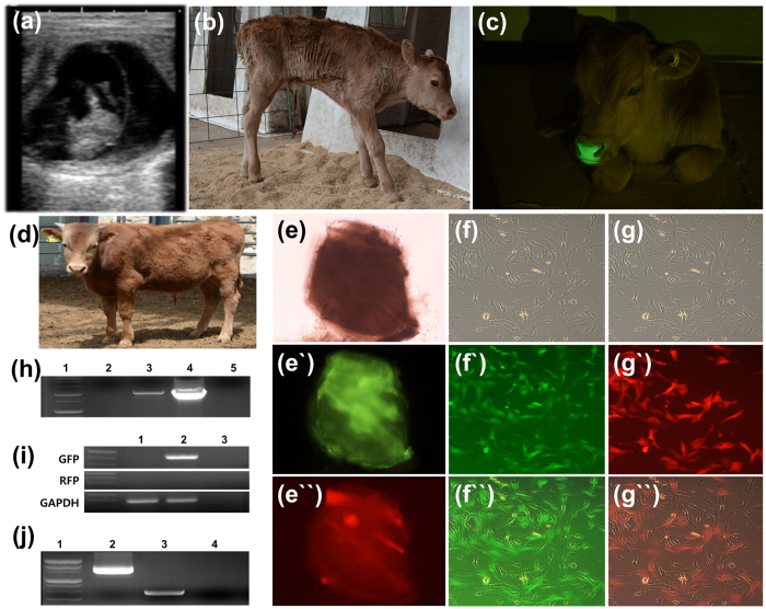
Figure 3. Birth of a transgenic (tg) cattle with the rox-GFP-rox-RFP gene via Piggybac (PB) and its analysis.
(a) After 45 days of embryo transfer, pregnancy was confirmed by ultrasonography. (b) The calf was delivered without assistant. (c) When ultraviolet light was exposed to nose of tg cattle, GFP expression was strongly observed. And the tg cattle grew up to 12 months old without any healthy issue (d). To determine GFP or RFP expression in a piece of tissue or primary skin cells via recombination, the tissue and cells were cultured and transfected with Dre recombinase mRNA by nucleofection ((e) a piece of tissue from tg cattle-brightness, (e`) before Dre recombinase transfection (GFP), (e``) after Dre recombinase transfection (RFP)). The primary skin cells from the tg cattle were isolated, cultured and transfected with Dre recombinase mRNA. Before transfection, only GFP expression was observed, RFP expression were observed via GFP gene excision by recombination ((f–f``) before transfection brightness, fluorescence, and merged, respectively; (g–g``) after transfection brightness, fluorescence, and merged, respectively). The transgene integration and recombination were confirmed by genomic DNA PCR ((h) 1: Molecular maker, 2: Wild type cattle, 3: Blood from tg cattle, 4: Positive control (DNAs), 5: Negative control) and RT-PCR ((i) 1: Wild type cattle, 2: cDNA from tg cattle, 3: Negative control). After Dre recombinase transfection, GFP excision was confirmed by genomic DNA PCR ((j) 1: Molecular marker, 2: Before transfection, 3: After transfection, 4: Negative control). Gel image was cropped and original image was seen in Supplementary Figure 8.

