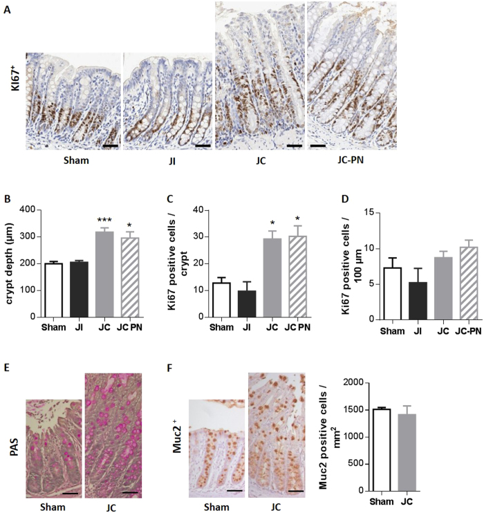
Figure 2. histological adaptation of the remaining colon mucosa after intestinal resection in rats.
(A) Representative photomicrographs of colon mucosa of Sham, IR jejuno-ileal (JI), IR jejuno-colonic (JC) and IR jejuno-colonic with PN (JC-PN) rats immunostained with an anti-Ki67 antibody. (B) Measurement of colon mucosa crypt depth in μm (at least 5 crypts analyzed by rat). (C) Quantification of Ki67 positive cells per crypt (at least 5 crypts analyzed by rat), (D) density of Ki67 positive cells expressed as number of Ki67 positive cells per 100 μm of crypt. (E,F) Representative photomicrographs of colon mucosa of sham and IR-JC rats stained with Periodic Acid Schiff (PAS) (E) and Representative photomicrographs of colon mucosa of sham and IR-JC rats immunostained with anti-Muc2 antibody (F) with quantification of Muc2 positive cells per area in mm2. Data are represented as mean ± SEM of n = 6 for sham, n = 4 to 6 for IR jejuno-ileal, n = 10 for IR jejuno-colonic, n = 5 to 6 for for IR jejuno-colonic with PN. *P < 0.05, **P < 0.01, vs sham-operated rats based on non-parametric Kruskal-Wallis test followed by Dunn’s adjusted multiple comparisons. Scale bar: 50 μm.

