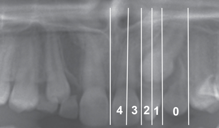
Figure 2.
Panoramic view illustrating reference lines of canine overlap (sectors) assigned to one of five categories: −1= distal to the normal position (in the premolar region), 0 = normal position (primary canine), 1 = distal to the long axis of the lateral incisor, 2 = mesial to the long axis of the lateral incisor, 3 = distal to the long axis of the central incisor, or 4 = mesial to the long axis of the central incisor.

